A recent study found that childhood cancers survivors are more than twice as likely to develop melanoma as adults, when compared to those who did not have childhood cancer.
The largest risk comes from receiving radiation and certain types of chemotherapy, which can affect skin cells even years down the road, making them more susceptible to skin cancer. In the study, patients were most likely to have melanoma on the torso, followed by the arm, leg and neck. These are the areas that were most likely to be radiated.
“Radiation destroys cancer cells by breaking the tumor’s DNA,” says Katherine Cools, M.D., a surgical oncologist at Henry Ford Health. “But sometimes healthy cells also get destroyed and the body can’t successfully repair itself.
"Childhood cancer survivors are a unique group – they are receiving tough treatments at a time when their bodies are undergoing rapid growth. The development of melanoma is probably due to the multiple hits their tissue took from treatment and the repair required to try to fix that area. The more times the body needs to repair itself, the more opportunity there is for mistakes to be made in the DNA – and for cancer to develop.”
Cancer Treatment & Melanoma Risk
The study examined two different chemotherapies called alkylating chemotherapy and doxorubicin, along with radiation. Researchers were able to identify the amount of each treatment that led to an increased risk of melanoma.
“Each dose of radiation is quantified in a unit called grays,” says Dr. Cools. “The study found that if the gray amount was greater than 40, a patient was more likely to develop melanoma in that area.
“For alkylating chemotherapy, doses are measured in what’s called ‘CED.’ If the CED was above 20,000, it led to an increased risk of melanoma. For doxorubicin, there was an increased risk of melanoma if they received it at all, regardless of the amount.”
Environmental & Genetic Risk Factors For Melanoma
There are also genetic components that increase someone’s risk of melanoma. For example, once you’ve had cancer, you are at a higher risk for getting another (unrelated) cancer.
“We also do know of a few genes that are associated with melanoma,” says Dr. Cools. “There is familial atypical mole melanoma syndrome (FAMMS) and the BRCA2 mutation (most commonly associated with breast cancer), along with the TP53 and RB1 mutations. People with a family history of melanoma are also at an increased risk of melanoma, along with those who are fair-skinned and have a lot of atypical moles.”
There are also environmental factors that increase the risk of melanoma, like UV exposure from the sun, tanning beds and even welding.
How To Reduce Your Risk Of Skin Cancer
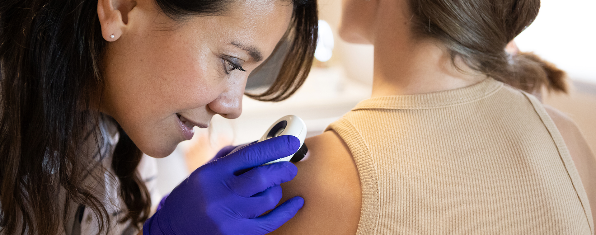
Currently, there isn’t skin cancer screening guidance that’s specific to childhood cancer patients. But if you had a substantial amount of radiation exposure, it is recommended to have annual skin checks, where your primary care doctor or dermatologist examines the entire surface of your skin for any unusual-looking moles that could signify cancer.
It is also recommended that everyone – regardless of their skin cancer risk – limit UV exposure as much as possible. Wear sunscreen and UV protective clothing and stay out of the sun between the hours of 10 a.m. and 4 p.m.

Melanoma Treatment At Henry Ford Health
“This study shows we should be screening childhood cancer survivors for melanoma,” says Dr. Cools. “As an adult, you might not know what treatments you received as a child, but your doctor can get an idea based upon the type of cancer you had. If we can get more of this type of information, we can hopefully screen childhood cancer survivors closely and let them know about their potential risk of melanoma. At a minimum, we can have them do self-checks for skin cancer.”
How to do a self-check? Scan your entire body for any unusual-looking moles. The guidelines for what’s considered unusual are:
- A: Asymmetry (one half of the mole is different from the other half)
- B: Border (the border of the mole is irregular)
- C: Color (the color of the mole differs from one spot to another)
- D: Diameter (the diameter is greater than 6 millimeters)
- E: Evolving (the mole has changed over time)
If you have a mole that checks any of the above boxes – or one you’re unsure about – reach out to your primary care doctor or dermatologist. The earlier cancer is spotted, the more treatable it is.
Reviewed by Katherine Cools, M.D., a surgical oncologist who sees patients at Henry Ford Cancer – Detroit and Henry Ford Medical Center – Bloomfield Township.


