Related News


COVID-19 Linked to Rare Form of Encephalitis
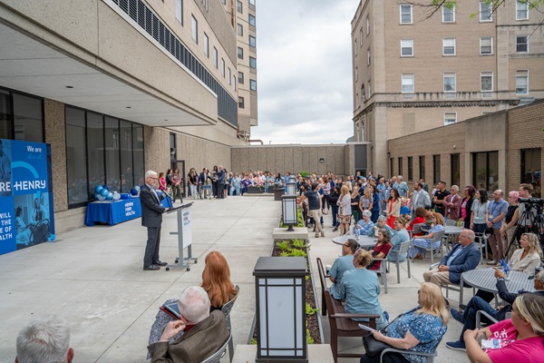
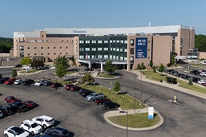
DETROIT – Henry Ford Health System is reporting an unusual case of encephalitis associated with COVID-19.
In a case report published online Tuesday in the journal Radiology, a team of doctors say a patient who tested positive for the coronavirus developed a case of acute necrotizing encephalitis, or ANE, a central nervous infection that mostly afflicts young children.
The 58-year-old female patient is hospitalized in serious condition.
This case report is believed to be the first published case highlighting the association between encephalitis and COVID-19.
“This is significant for all providers to be aware of and looking out for in patients who present with an altered level of consciousness. We need to be thinking of how we’re going to incorporate patients with severe neurological disease into our treatment paradigm,” says Elissa Fory, M.D. a Henry Ford neurologist who was part of the team of medical experts involved in making the diagnosis. “This complication is as devastating as severe lung disease.”
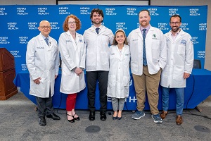
The team involved neuroradiologist Brent Griffith, M.D., infectious diseases physician Pallavi Bhargava, M.D., and neurologists Shaneela Malik, M.D. and Poonam Bansal, M.D.
Dr. Griffith, senior author of the published case report, says the paper shows “the important role that imaging can play in COVID-19 cases.”
ANE is a rare condition, particularly in the adult population, and is associated with poor clinical outcomes. It develops in response to other infections like influenza, chickenpox and enterovirus.
The patient had several days of fever, cough and muscle aches – symptoms consistent with COVID-19. On March 19, she was transported by ambulance to the emergency department and showed signs of confusion, lethargy and disorientation, Dr. Fory says. A flu test turned up negative but a rapid COVID-19 test, developed in-house by Henry Ford’s clinical microbiology lab, confirmed positive coronavirus.
When the patient remained lethargic, doctors ordered a repeat CT and MRI scans. The MRI scan identified abnormal lesions in both thalami and temporal lobes, parts of the brain that control consciousness, sensation and memory function. These scans confirmed doctors’ early suspicions.
“The team had suspected encephalitis at the outset, but then back-to-back CT and MRI scans made the diagnosis,” Dr. Fory says.
###
MEDIA CONTACT: David Olejarz / David.Olejarz@hfhs.org / 313.874.4094