Related News


Henry Ford Treats World’s First Patient Using New MRI-Guided Radiation Therapy that Simultaneously Tracks, Treats Tumor
DETROIT – The Henry Ford Cancer Institute has treated the world’s first cancer patient today with an advanced radiation therapy that uses an FDA-cleared real-time magnetic resonance imaging and linear accelerator delivery for more precise and accurate radiation treatment.
The MRIdian Linac by ViewRay, Inc. can treat cancer anywhere in the body. It is the world’s first and only FDA-cleared commercially available linear accelerator-based MRI-guided radiation therapy system that can image and treat patients simultaneously. The FDA cleared use of the company’s next generation model in February.
This next generation MRIdian Linac system is a novel radiation oncology technology that delivers:
- Confidence in accuracy and precision. Real-time high-quality MRI images and continuous soft-tissue imaging is done throughout the treatment automatically targeting the tumor and surrounding organs, ensuring the beam hits the targeted treatment area.
- Personalized treatment. By continuously observing and assessing the patient’s tumor and internal organs, clinicians can adapt in real-time, reshaping the dose to match each patient individually while on the table.
- Real-time imaging. When clinicians can clearly see the target and watch where the radiation is being delivered, they are better able to adapt to natural movement of the patient’s anatomy. Doctors previously did not have the tools to see organs in real time to make changes during radiation therapy.
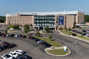
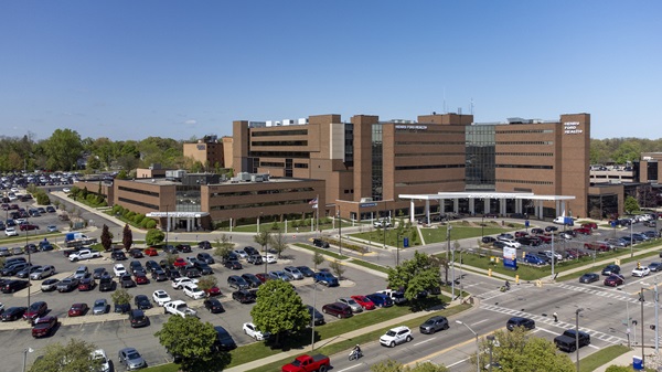
The first clinical MRIdian Linac system is available at Henry Ford Medical Center-Cottage in Grosse Pointe Farms. The first patient in the world was treated there Wednesday, July 19 for prostate cancer. Using this new technology radiation oncologists were not only able to see the exact location of the prostate in real-time as it was being treated, but also the neighboring normal structures, including the bladder and rectum.
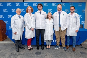
“This is an incredibly exciting advancement in our field that is unlike anything else in the world to treat cancer patients,” says Benjamin Movsas, M.D., chair of the Department of Radiation Oncology at the Cancer Institute and a national expert.
“Being able to continuously see both the tumor and surrounding organs means our team can accurately align the tumor to the treatment beams and make adjustments in real time to avoid sensitive internal structures. For radiation oncologists, this exciting technology finally ‘takes off the blindfold,’ and allows us to see during treatment what we could not previously visualize. This is a major leap forward that will benefit cancer patients.”
The new system can be used to treat all types of cancers anywhere in the body, and is especially beneficial for tumors where there is typically movement during treatment, including tumors in the liver, pancreas, adrenal and lung. Other types of cancer this advanced system will deliver a new level of care to include breast, prostate, kidney, and gynecologic cancers, among others.
Indrin Chetty, Ph.D., division head of Physics in Radiation Oncology, says the MRIdian Linac system further enhances radiation therapy with high-quality soft tissue images that make it possible to precisely align and adapt to changes, as well as shrink margins while increasing doses.
“For a pancreatic cancer patient, for example, we can now see the exact shape and location of the tumor as well as nearby critical structures to avoid the stomach, liver and small intestine throughout the treatment,” he says. “Now we can account for shape and position of the tumor to its surrounding organs to safely deliver ultra-high doses offering a better chance for local control.”
Over the years, technology has improved the accuracy of radiation treatment while protecting surrounding healthy tissue. However, trying to accommodate for the natural movement of a tumor and the body’s internal organs during treatment has been elusive.
“We believe our system will lead to a new standard of care in radiation oncology,” says Chris Raanes, president and CEO of ViewRay. “With ViewRay’s first generation MRIdian system treating nearly all cancer types, clinicians saw for the first time how much tumors and organs move and change shape during the course of treatment.”
The ViewRay system combines the effectiveness of MRI, which produces high-quality images of organs and structures inside the body, with a linear accelerator to map out a therapy plan and deliver radiation at the intended target, while allowing for refinements to be made in real-time during treatment. The result is a more accurate, precise treatment.
“Our Department of Radiation Oncology has a long tradition of expertise using the most advanced radiation technologies, and the ViewRay system offers our patients another optimal treatment for achieving the best care possible,” says Steven Kalkanis, M.D., medical director of the Cancer Institute.
Nearly two-thirds of all cancer patients in the United States will receive some form of radiation therapy during their course of treatment. In use for more than 100 years, radiation therapy damages the genetic material in cancer cells and helps to prevent the cells from growing and spreading. Treatments are delivered using specialized procedures to enhance their safety and effectiveness.
For patients interested in learning more about ViewRay: Call (888) 777-4167 or visit www.henryford.com/preciseradiation
About the Henry Ford Cancer Institute
The Henry Ford Cancer Institute is one of the largest cancer programs in Michigan, providing care at four hospitals and four outpatient facilities throughout southeast Michigan. Treatment for the most complex or rare cancers and the Institute’s extensive cancer research program is anchored by Henry Ford Hospital. For more information, visit www.henryford.com/cancer.
MEDIA CONTACT: Krista Boyer