Division of Ultrasound
The Henry Ford Hospital Department of Emergency Medicine values the key role Point of Care Ultrasound plays in the delivery of high-quality, cutting-edge emergency care. The Department of Emergency Medicine Division of Ultrasound established the first Emergency Medicine Clinical Ultrasound Fellowship Program in the state of Michigan beginning in July of 2010.
For more information of our Clinical Ultrasound Fellowship please click here.
Resident ultrasound education
 Resident ultrasound training is based on the ACEP imaging compendium and recently published ultrasound milestones. Resident ultrasound training is based on the ACEP imaging compendium and recently published ultrasound milestones. Residents are required to archive 150 scans before graduation, using our wireless image archiving system (Q-path E).
Our ultrasound faculty and the ultrasound fellow are readily available to assist residents in the ED with scanning and providing immediate feedback. Residents also receive feedback on studies they perform via our Quality Assurance system. The training our residents receive meet all the basic requirements as recommended by the Residency Review Committee (RRC), and the ACGME. By the completion of the program, residents will be able to integrate the latest Point of Care Ultrasound (POCUS) techniques into their daily practice.
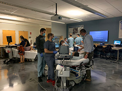
We have a longitudinal curriculum where residents develop their ultrasound skills throughout their three years or training. Below is a description of the resident ultrasound curriculum.
Interns
The emphasis during the first year is on building a strong foundation for residents to develop the necessary competencies in all aspects of POCUS. Residents become familiar with the ultrasound user interface and perform common ED ultrasound exams.
Ultrasound training during this year is comprised of the following:
- Orientation Month: All residents taking a 2-day POCUS course which includes didactics and hands-on practice sessions using live models and phantoms. This session introduces residents to all aspects of ultrasound. Residents will also complete their Ultrasound-Guided Central Venous Access training during this month.
- Ultrasound Rotation: This is a one-week rotation of Ultrasound scanning in the Emergency Department. During this week, residents have one-on-one scanning sessions with the Ultrasound team-
- Sonosim: Asynchronous ultrasound learning modules. Through the rest of the year, there are ultrasound objectives developed for outside rotations.
- Simulation Center Didactics: Every two months we have hands-on scanning sessions in the simulation center using live models as a part of DEM Grand Rounds.
Additionally, residents scan during their clinical shifts while rotating in the ED and can participate as teaching assistants during the numerous Wayne State University School of Medicine ultrasound training sessions.
Junior residents
The second year is dedicated to a systems-based approach to ultrasound training.
- Scan Shifts: During each block rotation in the Department of Emergency Medicine, residents are given on Scan Shift. These shifts include hands-on bedside scanning with Ultrasound fellow and faculty, QA of studies, completion of asynchronous learning modules, and completion of procedures in the Emergency Department which use Ultrasound. Common procedures performed during these shifts include peripheral IVs, arterial lines, paracentesis, thoracentesis, central venous access (internal jugular vein), arthrocentesis, and nerve blocks.
- Sonosim: Throughout the rest of the year, there are ultrasound objectives developed for outside rotations.
- Simulation Center Didactics: Every two months we have hands-on scanning sessions in the simulation center using live models as a part of DEM Grand Rounds. During these sessions we deepen the resident’s understanding of the pathology that can be identified using ultrasound, focusing on musculoskeletal, echocardiography/hemodynamics, abdominal/trauma, procedural, and obstetrics.
Additionally, while on ICU rotations, residents receive additional training in critical care applications of POCUS from the Critical Care Ultrasound faculty. The juniors also have the opportunity to participate during the Wayne State University School of Medicine training sessions where they serve as instructors to medical students during small group training sessions.
Senior residents
By third year, residents are complete the required 150 ultrasound scans required for resident training. Many of our residents also complete additional scans to fulfill the requirement for ultrasound credentialing. During their PGY3 year, seniors receive ultrasound education in the following manner:
- Simulation Center Didactics: Our seniors also participate every two months in hands-on scanning sessions in the simulation center using live models as a part of DEM Grand Rounds. During these sessions we teach advanced ultrasound skills and deepen our Senior residents’ understanding of the pathology that can be identified using ultrasound, focusing on musculoskeletal, echocardiography/hemodynamics, abdominal/trauma, procedural, and obstetrics. For the PGY3 curriculum we begin to incorporate Fellowship-level education.
- Ultrasound Elective: Residents can complete a 4-week elective during their senior year. During this block, our residents have the opportunity to participate in ongoing research projects, develop their teaching skills by working with more novice sonographers, and learn about advanced applications of ultrasound including echocardiography and hemodynamic assessment of critically ill patients.
- Ultrasound teaching: Senior residents enhance their ultrasound skills by directly supervising medical students who are assigned to work with them during scheduled ED shifts while rotating in the Emergency Department.
- Seniors can participate as instructors during the WSUSOM training sessions.
Medical student training
The Henry Ford Hospital has a unique affiliation with the WSUSOM to train medical students in the use of bedside ultrasound through a partnership with General Electric (GE). This partnership was made possible through a $5M grant to the medical school in 2006 with the goal of incorporating ultrasound education into the medical school curriculum at the largest single campus medical school in the nation. A longitudinal curriculum has been developed for medical student training across all 4 years of their medical education. This has been a very successful program and has given residents, fellows and faculty excellent teaching opportunities.
Henry Ford Ultrasound University:
The HFH Ultrasound University was established in 2009, with the goal of promoting the widespread use of ultrasound by acute care practitioners in various clinical settings, and to serve as a resource of excellence in patient care and safety standards. Many formal ultrasound courses with CME credits are offered throughout the year, including special courses designed to train residents in specialties such as Urology, General Surgery, Internal Medicine, and Critical Care.
EM residents and fellows have opportunities to participate in these courses as learners and instructors.
Ultrasound equipment
The DEM currently has nine ultrasound systems, including:
- Three SonoSite X-Porte
- Three Sonosite S2s
- Two Mindray M9s
- 1 GE VividQ which has Strain capability.
All systems are equipped with multiple transducers for all the various EM ultrasound applications, including endocavitary and transesophageal probes.
The department also purchased several Butterfly iQ probes for resident and fellow use. All of the systems have been set up for wireless image archiving using Q-path E, which allows for easy storage of images, provides statistical analysis, facilitates immediate feedback of images, and helps streamline the QA process. Additionally, we have the capability to bill for scans which are performed by credentialed physicians. These scans automatically upload to the PACS system in EPIC and are viewable by all and are a part of the patient’s medical record.
TEE in the ED
The HFH ED Resuscitative Transesophageal Echocardiography program, with the support of the HFH Cardiothoracic Division of Anesthesia, was established in April 2022. This program has been successfully guiding cardiac arrest management since its inception. We currently have two TEE probes and are working on increasing the number of credentialed providers.